Detection and Imaging of Protein Microcrystals using SONICC®
Detection and Imaging of Protein Microcrystals using SONICC®
Authors: Ellen Gualtieri, Eric Zhao, Lance Ramsey
About SONICC®
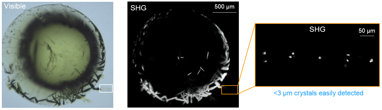
SONICC, Second Order Non-linear Imaging of Chiral Crystals, uses a femtosecond pulsed laser to exploit the frequency-doubling effect found in the majority of protein crystals, and produces high-contrast images with negligible background signal. SONICC has two imaging methods, Second Harmonic Generation (SHG) and Ultraviolet Two-Photon Excited Fluorescence (UV-TPEF). The SHG channel probes crystallinity, and the UV-TPEF channel is specific to proteinaceous samples.
SONICC Imaging
SONICC can be used to automatically screen all types of crystallization plates to positively identify both large and small crystals. It is especially useful for detecting crystals buried in precipitate or turbid matrices, such as lipidic cubic phase (LCP). LCP is often used to crystallize membrane proteins with initial screens resulting in small crystals that are not easily identified in the turbid LCP matrix. SONICC is particularly well suited for imaging in LCP as it can detect small crystals even in the presence of a turbid matrix. Automated imaging tools such as drop location and auto focus are both used to quickly and accurately find the LCP drop. The accompanied visible imaging technique of crossed-polarized imaging offers complimentary information all with extremely fast imaging times.

Microcrystalline Detection
SONICC can detect crystals <400 nm3 and therefore is well suited for imaging protein crystals that are not easily imaged with conventional techniques. Recent advances in nanocrystallography by Fromme Lab at Arizona State University demonstrated the need for an alternate technique to characterize protein samples containing sub-micron crystals. In collaboration with Ross at ASU, they have used SONICC to image protein crystals in microfluidic channels.
Easily Find Hits
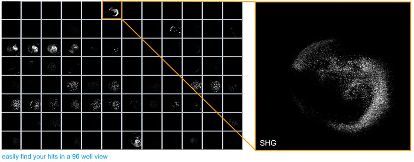
The stark black and white images makes it easy to identify positive hits, even microcrystals, in a 96 well view reducing the amount of time spent on looking at images.
References
1. Kissick, D. J.; Gualtieri, E. J.; Simpson, G. J.; Cherezov, V., Nonlinear optical imaging of integral membrane proteins in lipidic mesophases. Anal. Chem. 2010, 82(2), 491-497. 8. Wampler, R. D.; 2.
2. Kissick, D. J.; Dehen, C. J., Gualtieri, E. J.; Grey, J. L.; Wang, H.; Thompson, D. H.; Cheng, J.; Simpson, G.J., Selective detection of protein crystals by second harmonic microscopy. Journal of American Chemical.
3. The detection and subsequent volume optimization of biological nanocrystals.Struct. Dyn. 2, 041710 (2015)