SONICC® (Second Order Nonlinear Imaging of Chiral Crystals) is a novel imaging technique that offers unprecedented sensitivity in quantifying small molecule crystallinity. SONICC can be used to detect concentrations of crystalline APIs down to 0.03% in amorphous solid dispersions. Analysis is fast (< 1 second), no sample preparation is required, and can be performed on final form with excipients present.
Amorphous solid dispersions
The bioavailability of the active pharmaceutical ingredient (API) in a drug is typically a strong function of the solubility of the API molecule itself. When polymorphic forms of this molecule are present, the most stable one is usually crystalline. Unfortunately, this same crystalline polymorph is also generally the least soluble. In such cases it becomes desirable to develop formulations based on the amorphous form of the API, with stabilization obtained by the addition of precise combinations of additives. Polymers and other excipients are used in varying amounts to create stable formulations that retard the conversion of the more soluble amorphous API to its less soluble crystalline form. For purposes of optimization, these formulations are subjected to various stresses (temperature, humidity. etc.) to simulate accelerated aging while being monitored to ensure they remain amorphous, preserving their dissolution characteristics. Common techniques for analyzing the state of the API within a formulation include x-ray powder diffraction, birefringence, Raman microscopy and calorimetry. These techniques each exhibit their own trade-offs between sensitivity and selectivity, with X-ray diffraction generally considered the most powerful; detection limits down to 1-2% crystallinity are routinely observed. Polarized light microscopy (PLM) offers lower detection limits down to the single particle regime, however it lacks selectivity. All crystals, including excipient crystals will be detected with PLM making it not useful to analyze formulations in tablet dosage form.
Unprecedented sensitivity with SONICC
A new characterization technique called SONICC (Second Order Nonlinear Optical Imaging of Chiral Crystals), recently developed by Prof. Garth Simpson at Purdue University and distributed under exclusive license by Formulatrix, achieves unprecedented sensitivity to chiral crystals with a currently demonstrated Limit Of Quantitation (LOQ) down to 0.03% crystallinity.1,2 SONICC uses multi-photon microscopy to selectively detect chiral crystals with no interference from amorphous or achiral materials. SONICC relies on Second Harmonic Generation (SHG), where two low-energy photons combine under intense electric fields to form a higher energy photon. This process only occurs in noncentrosymmetric ordered crystals. Thus the signal is generated only in the presence of chiral crystals; background signal is extremely low, resulting in excellent limits of detection for chiral crystalline small molecules.
SONICC for absolute quantitation
Quantitation of crystalline API within a particular formulation can provide insight into the stability of the formulation and help determine its effectiveness in stabilizing the API. Using calibration standards, SONICC can determine the concentration of crystalline material in an unknown sample. For proof of concept, tablets of amorphous griseofulvin and copovidone were spiked with varying amounts of crystalline griseofulvin. A calibration curve of SONICC response versus spiked amount was plotted and fit with a straight line (Figure 1). A test sample was prepared at 2% crystalline material by weight and analyzed with SONICC in triplicate. Using the calibration curve shown in Figure 1, the test sample was determined to be 1.6% +/- 0.3% crystallinity. Preparation of a sample exactly to 2% was not trivial and is the largest source of error in these measurements. A SHG image of one of the standard tablets is shown in Figure 4. It is evident from the microscopy image that the crystalline griseofulvin is not evenly distributed in the tablet, thus resulting in error for the analysis of the test sample.
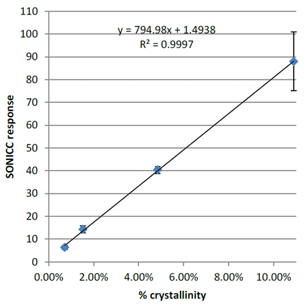
Figure 1. Calibration curve of SONICC response versus % crystallinity for amorphous griseofulvin/copovidone tablets with spiked known amounts of crystalline griseofulvin.
An order of magnitude improvement in LOQ over Powder X-Ray Diffraction (PXRD) was demonstrated in a study where samples of pure crystalline griseofulvin were analyzed with both SONICC and XRD after being cryomilled for increasing amounts of time. As can be seen in Figure 2, after 1 hour of cryomilling, PXRD could no longer detect any crystalline material, whereas after 3 hours, SONICC still detected 0.05% crystalline material present.2 This order of magnitude improvement in the LOQ using SONICC provides the ability to assess samples with low amounts of crystallinity and enables the analysis of samples with low drug loading.
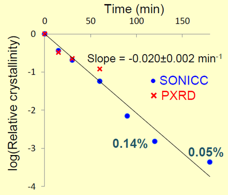
Figure 2. Relative crystallinity versus cryomilled time of crystalline griseolfulvin sample.2
SONICC for relative quantitation
It is not always feasible or necessary to obtainabsolute quantitation of the extent of crystallization of an API in a sample. Often a
relative difference between samples is sufficient in evaluating various formulations. For example, a drug formulation (non-disclosed for proprietary reasons) was analyzed with different drug loading concentrations under stressed and unstressed conditions. This particular case study was for samples with very low drug loading (0.03% and 3.2% drug concentration) and would not have been easily assessed with conventional techniques like PXRD. As seen in Figure 3, SONICC could easily identify the relative differences in percent crystallinity between the various formulations. Even with the sample containing only 0.03% API, SONICC could still discern between the stressed and unstressed samples. SONICC also indicated that the 3.2%
concentration unstressed sample had significant crystalline material present compared to the placebo and 0.03% unstressed samples.

Figure 3. Relative SHG response for drug formulation with varying amounts of drug loading (0.03% and 3.2%) under stressed and unstressed conditions.
SONICC provides spatial distribution information
The fact that SONICC is an imaging technique also provides helpful information regarding the spatial distribution of crystalline drug in a sample. This can be especially useful when assessing various mixing techniques. For example, a powder sample was prepared with amorphous griseofulvin, copovidone and small amounts of crystalline griseofulvin. A SONICC image can be seen in Figure 4, where a large patch of crystalline material can be identified in the dispersion. 2mm square images with 3um x,y resolution are acquired in 500 ms. Because SONICC is a multi-photon technique, depth penetration up to 500 μm can be achieved with ~30 μm resolution in z. 3-D renderings of a griseofulvin/copovidone tablet are shown in Figure 5. This is especially useful for analyzing tablets with coatings. SONICC can penetrate through the coatings and give information as to how the drug is localized and whether or not it is in a crystalline or amorphous form.
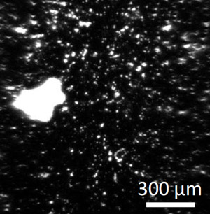
Figure 4. SONICC image of crystalline griseofulvin in an amorphous griseofulvin/copovidone tablet.
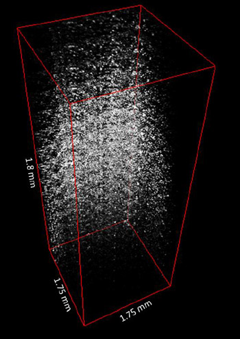
Figure 5. SONICC 3-D rendering of crystalline griseofulvin in an amorphous griseofulvin/copovidone tablet.
SONICC can also be used in a mode where it detects UV excited fluorescence. This is especially useful for visualizing where the API, in either crystalline or amorphous form, is located in the formulation. The UV fluorescence mode is specific to molecules, like APIs that fluoresce but is not selective to crystallinity. Together, the UV mode with the crystal specific SHG mode can show where the drug is localized and what form it is in. A multiphoton UV fluorescence image of an amorphous griseofulvin/copovidone tablet can be seen in Figure 6.
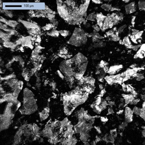
Figure 6. UV two photon excited fluorescence image of an amorphous griseofulvin/copovidone tablet.
Conclusion
The extremely low level of detection and selectivity offered by SONICC enables scientists to learn more about their formulations. SONICC provides a means of analyzing formulations with low drug loading that could otherwise not be studied with conventional techniques. SONICC offers the sensitivity and imaging information that is obtained with polarized light microscopy while still providing selectivity to the API. The ability to conduct meaningful assessments at significantly accelerated time points is envisioned. Access to SONICC instruments and services are available exclusively through Formulatrix.
To learn more about SONICC for API,
please email info@formulatrix.com
References
1. Wanapun, D.; Kestur, U. S.; Simpson, G. J.; Taylor, L. “Selective Detection and Quantitation of Organic Molecule Crystallization by Second Harmonic Generation Microscopy” Accelerated Article, Anal. Chem. 2010, 82, 5425–5432
2. Wanapun, D., Kestur, U. S., Kissick, D. J., Taylor, L. S., and Simpson, G. J. “ Single particle nonlinear optical imaging of trace crystallinity in an organic powder.” Anal. Chem.2011, 83(12), 4745-51.
Further Reading
Kestur, U. S., Wanapun, D., Toth, S.J. , Wegiel, L. A., Simpson, G. J. and Taylor, L. S. “Nonlinear Optical Imaging for Sensitive Detection of Crystals in Bulk Amorphous Powders.” Journal of Pharmaceutical Sciences, 2012.
Toth, S.J., Madden, J. T., Taylor, L. S., Marsac, P., and Simpson, G. J. “Selective Imaging of Active Pharmaceutical Ingredients in Powdered Blends with Common Excipients Utilizing Two-Photon Excited Ultraviolet-Fluorescence and Ultraviolet-Second Order Nonlinear Optical Imaging of Chiral Crystals.” Analytical Chemistry 2012, 84, 5869-5875