Image Quality with Highest Resolution Available
Fixed Objective Option
Two objectives are available for imaging protein drops. The large field of view objective is used for locating drops with two high magnification objectives available for high-resolution imaging. The software automatically determines the best objective to use based on the size of the drop. This option is available on any Rock Imager® system upon request.
Continuous Zoom Optics
Infinite zoom values are available up to 12x magnification allowing you to zoom in exactly to drop sizes and Regions of Interest (ROI). This option is available on any Rock Imager system, except the Rock Imager 1, upon request.


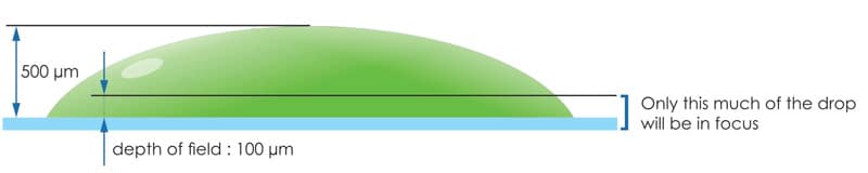
Extended Focus Imaging (EFI)
Maintain High Resolution and High Depth of Field
High resolution means that you have a lower depth of field when imaging drops, so the entire drop will not be in focus at the same time. To achieve high optical resolution and high depth of field, Rock Imager captures several images at different Z-heights (slices) and computes an image that contains the best-focused pixels from each of the slices. This is called Extended Focus Imaging.
More Tools for Better Imaging Results
Formulatrix® offers the greatest functionality of any automated visible imager on the market. Drop location, auto-focus, continuous zoom optics and fast color imaging are combined together to create scientific publication-worthy images on a routine basis. There is no need to go back to drops to then tweak settings to get a publication quality image. Extremely fast stage movement and image acquisition allows you to obtain high-resolution images for every single plate and drop imaged.
High Resolution 12x Zoom
High resolution 12x variable zoom offers you an infinite number of magnifications for manual or automated imaging at the perfect zoom needed.
- High NA (numerical aperture) objective provides sharp high contrast images
Kohler Illumination with Motorized Aperture
Rock Imager uses Kohler Illumination, the same light source found in compound microscopes. This arrangement uses lenses to focus light on the sample. A motorized aperture in the light path allows this light to be morphed from a cone of light. A cone of light produces images with the least amount of shadowing. A column of light produces images with the highest contrast.
- No sample heating due to IR filter and remote location of lamp
- Motorized control of aperture automates contrast adjustment
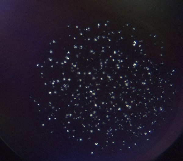
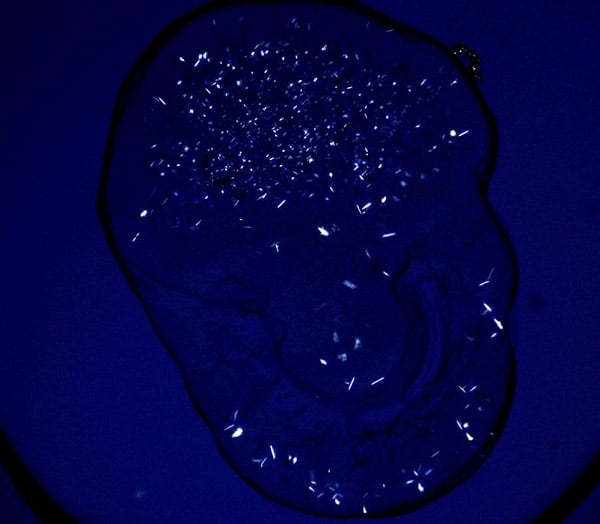
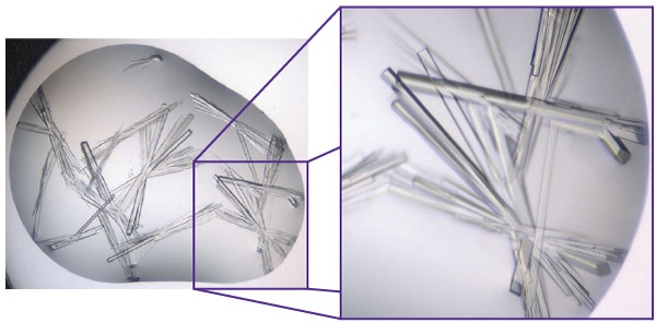
See Thin Crystals and Capture More Detail
Birefrigent imaging makes it possible to visualize thin crystals that may otherwise be missed. It is also useful in distinguishing between salt and protein crystals in some cases. Crossed-polarized imaging is especially useful in LCP crystallization setups where the crystals are so small. High contrast images are generated with dark backgrounds (99.95% extinction ratio).
When not performing birefringent imaging, the polarizers in the optical pathway can cause unwanted shadowing and drop discoloration due to plate and plate seal interference. To prevent this interference, the top polarizer is automatically removed from the light path when not in use resulting in crystal clear brightfield images.
Imaging Made Easy
Auto-focus
Auto-focus is used to accurately find your drop in the z-direction. This is especially useful for imaging hanging drops on greased cover slides or when zooming into a region of interest. Auto-focus works by capturing a stack of images at 30 frames per second and computing a focus curve. The imager can then jump to the best focus image or determine the best z range to capture slices to create an extended focus image.
Auto-exposure
Auto-exposure finds the correct settings for both the illumination intensity and the camera exposure time to maximize image brightness without overexposure.
Automatic Drop Location
Rock Imager has a fast, robust drop location algorithm that locates drops in three dimensions (X, Y, & Z). Once the drop is located, the zoom and X, Y position is adjusted to the center of the drop in the screen, providing maximum optical resolution. A specialized LCP drop location algorithm detects the precipitant drop and lipid drop, zooming in on the lipid drop for high high-resolution.
- Over 99% effective
- Motorized condenser is used to shadow drops making location extremely reliable
- Immune to defects such as small specs of dirt, condensation, or satellite drops
- Multiple drops can be located on a single coverslide
Found Something Interesting? Take a Better Look
Region of Interest (ROI)
In Rock Maker, you can draw a box on your image specifying an area to zoom into. The next time the plate is imaged, Rock Imager will automatically zoom into the area specified to capture a high-resolution image of the region of interest.
- Maximize the optical and digital resolution for a region
- Get detailed images of regions of interest without manual intervention
FAQs
What is visible light imaging for protein crystallization and how does it work?
Visible light imaging captures images using the visible light spectrum (400 to 700 nm wavelength). Techniques such as bright-field, dark-field, and phase-contrast microscopy utilize visible light for imaging. Visible light imaging works by illuminating crystallization drops with white light and capturing the reflected or transmitted light using a high-resolution camera.
How does visible light imaging capture high-resolution images of protein crystallization drops?
Visible light imaging uses visible light and precision optics to produce detailed images of crystallization drops. White light either passes through or reflects off the drop, and a high-quality camera captures the image through a microscope objective. Differences in how crystals, precipitates, and the surrounding solution scatter or reflect light create contrast, making crystal features clearly visible. In automated systems like Rock Imager, consistent lighting, focus, and positioning ensure reproducible, high-resolution images across all plates.
What microscopy techniques (bright-field, dark-field, phase contrast, polarized light) are used in visible light protein imaging?
All these microscopy techniques—bright-field, dark-field, phase contrast, and polarized light—are utilized in visible light imaging to enhance the visualization of protein crystals. Each technique offers unique advantages.