2020
Towards an Optimal Sample Delivery Method for Serial Crystallography at XFEL
Robert KY Cheng. Crystals 2020, 10(3), 215; https://doi.org/10.3390/cryst10030215
Received: 28 February 2020 / Revised: 15 March 2020 / Accepted: 17 March 2020 / Published: 19 March 2020
Preventing Bio-Bloopers and XFEL Follies: Best Practices from your Friendly Instrument Staff
Christopher Kupitz and Raymond G. Sierra. Crystals 2020, 10(4), 251; https://doi.org/10.3390/cryst10040251
Received: 5 March 2020 / Revised: 24 March 2020 / Accepted: 24 March 2020 / Published: 27 March 2020
Membrane Protein Preparation for Serial Crystallography Using High-Viscosity Injectors: Rhodopsin as an Example
Part of the Methods in Molecular Biology book series (MIMB, volume 2127)
2019
Strategies for sample delivery for femtosecond crystallography
I. Martiel, H. M. Müller-Werkmeister and A. E. Cohen. Acta Cryst. (2019). D75, 160-177 https://doi.org/10.1107/S2059798318017953
Sample Delivery Media for Serial Crystallography
Ki Hyun Nam. Int. J. Mol. Sci. 2019, 20(5), 1094; https://doi.org/10.3390/ijms20051094. Received: 15 February 2019 / Revised: 27 February 2019 / Accepted: 27 February 2019 / Published: 4 March 2019
Successful sample preparation for serial crystallography experiments
John H. Beale, Rachel Bolton, Stephen A. Marshall, Emma V. Beale, Stephen B. Carr, Ali Ebrahim, Tadeo Moreno-Chicano, Michael A. Hough, Jonathan A. R. Worrall, Ivo Tews and Robin L. Owen.
Sample delivery for serial crystallography at free-electron lasers and synchrotrons
M. L. Grünbein and G. Nass Kovacs. Acta Cryst. (2019). D75, 178-191
https://doi.org/10.1107/S205979831801567X
Visualization of protein crystals by high-energy phase-contrast X-ray imaging
M. Polikarpov, G. Bourenkov, I. Snigireva, A. Snigirev, S. Zimmermann, K. Csanko, S. Brockhauser and T. R. Schneider. Acta Cryst. (2019). D75, 947-958
https://doi.org/10.1107/S2059798319011379
Membrane protein megahertz crystallography at the European XFEL
Gisriel, C., Coe, J., Letrun, R. et al. Membrane protein megahertz crystallography at the European XFEL. Nat Commun 10, 5021 (2019) doi:10.1038/s41467-019-12955-3
Pembrolizumab microgravity crystallization experimentation
Reichert, P., Prosise, W., Fischmann, T.O. et al. Pembrolizumab microgravity crystallization experimentation. npj Microgravity 5, 28 (2019) doi:10.1038/s41526-019-0090-3
2018
Supersaturation-controlled microcrystallization and visualization analysis for serial femtosecond crystallography
Lee, D.B., Kim, J., Seok, J.H. et al. Supersaturation-controlled microcrystallization and visualization analysis for serial femtosecond crystallography. Sci Rep 8, 2541 (2018) doi:10.1038/s41598-018-20899-9
2017
Serial millisecond crystallography of membrane and soluble protein microcrystals using synchrotron radiation
Martin-Garcia, J. M., Conrad, C. E., Nelson, G., Stander, N., Zatsepin, N. A., Zook, J., Zhu, L., Geiger, J., Chun, E., Kissick, D., Hilgart, M. C., Ogata, C., Ishchenko, A., Nagaratnam, N., Roy-Chowdhury, S., Coe, J., Subramanian, G., Schaffer, A., James, D., Ketwala, G., Venugopalan, N., Xu, S., Corcoran, S., Ferguson, D., Weierstall, U., Spence, J. C. H., Cherezov, V., Fromme, P., Fischetti, R. F. & Liu, W. (2017). IUCrJ 4, 439-454.
XFELs for structure and dynamics in biology
Spence, J. C. H. (2017). IUCrJ 4, 322-339.
Locating and Visualizing Crystals for X-Ray Diffraction Experiments
Becker M., Kissick D.J., Ogata C.M. (2017) Locating and Visualizing Crystals for X-Ray Diffraction Experiments. In: Wlodawer A., Dauter Z., Jaskolski M. (eds) Protein Crystallography. Methods in Molecular Biology, vol 1607. Humana Press, New York, NY
Structures of riboswitch RNA reaction states by mix-and-inject XFEL serial crystallography
Nature 541, 242–246 (12 January 2017)
doi:10.1038/nature20599
2016
Preparation and Delivery of Protein Microcrystals in Lipidic Cubic Phase for Serial Femtosecond Crystallography
Ishchenko A, Cherezov V, Liu W. Preparation and Delivery of Protein Microcrystals in Lipidic Cubic Phase for Serial Femtosecond Crystallography. Journal of Visualized Experiments : JoVE. 2016;(115):54463. doi:10.3791/54463.
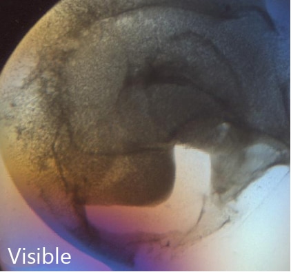
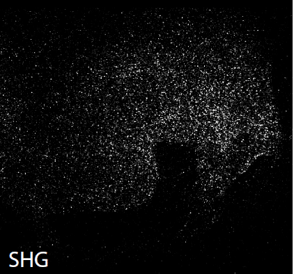
Nonlinear Optical Characterization of Membrane Protein Microcrystals and Nanocrystals
Newman J.A., Simpson G.J. (2016) Nonlinear Optical Characterization of Membrane Protein Microcrystals and Nanocrystals. In: Moraes I. (eds) The Next Generation in Membrane Protein Structure Determination. Advances in Experimental Medicine and Biology, vol 922. Springer, Cham
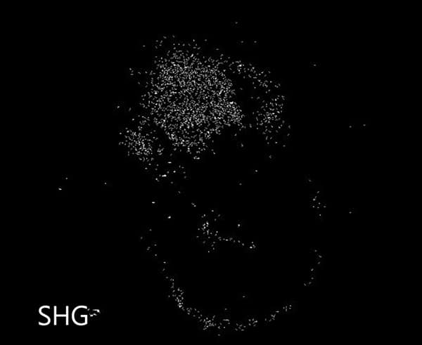
Second harmonic generation correlation spectroscopy for characterizing translationally diffusing protein nanocrystals
Dow XY, Dettmar CM, DeWalt EL, et al. Second harmonic generation correlation spectroscopy for characterizing translationally diffusing protein nanocrystals. Acta Crystallographica Section D, Structural Biology. 2016;72(Pt 7):849-859. doi:10.1107/S205979831600841X.
Serial femtosecond crystallography datasets from G protein-coupled receptors
Scientific Data 3, Article number: 160057 (2016)
doi:10.1038/sdata.2016.57
Guiding synchrotron X-ray diffraction by multimodal video-rate protein crystal imaging
Newman JA, Zhang S, Sullivan SZ, et al. Guiding synchrotron X-ray diffraction by multimodal video-rate protein crystal imaging. Journal of Synchrotron Radiation. 2016;23(Pt 4):959-965. doi:10.1107/S1600577516005919.
Transmission electron microscopy for the evaluation and optimization of crystal growth
Stevenson HP, Lin G, Barnes CO, et al. Transmission electron microscopy for the evaluation and optimization of crystal growth. Acta Crystallographica Section D, Structural Biology. 2016;72(Pt 5):603-615. doi:10.1107/S2059798316001546.
A pipeline for structure determination of in vivo-grown crystals using in cellulo diffraction
Boudes M, Garriga D, Fryga A, Caradoc-Davies T, Coulibaly F. A pipeline for structure determination of in vivo-grown crystals using in cellulo diffraction. Acta Crystallographica Section D, Structural Biology. 2016;72(Pt 4):576-585. doi:10.1107/S2059798316002369.
2015
Microfluidic sorting of protein nanocrystals by size for X-ray free-electron laser diffraction
Structural Dynamics 2, 041719 (2015); https://doi.org/10.1063/1.4928688
Serial femtosecond X-ray diffraction of enveloped virus microcrystals
Structural Dynamics 2, 041720 (2015); https://doi.org/10.1063/1.4929410
Batch crystallization of rhodopsin for structural dynamics using an X-ray free-electron laser
Wu W, Nogly P, Rheinberger J, et al. Batch crystallization of rhodopsin for structural dynamics using an X-ray free-electron laser. Acta Crystallographica Section F, Structural Biology Communications. 2015;71(Pt 7):856-860. doi:10.1107/S2053230X15009966.
Serial femtosecond crystallography: the first five years
Schlichting, I. (2015). IUCrJ 2, 246-255.
The detection and subsequent volume optimization of biological nanocrystals
Structural Dynamics 2, 041710 (2015); https://doi.org/10.1063/1.4921199
A comprehensive review of the lipid cubic phase or in meso method for crystallizing membrane and soluble proteins and complexes
Caffrey M. A comprehensive review of the lipid cubic phase or in meso method for crystallizing membrane and soluble proteins and complexes. Acta Crystallographica Section F, Structural Biology Communications. 2015;71(Pt 1):3-18. doi:10.1107/S2053230X14026843.
Reliably distinguishing protein nanocrystals from amorphous precipitate by means of depolarized dynamic light scattering
Schubert, R., Meyer, A., Dierks, K., Kapis, S., Reimer, R., Einspahr, H., Perbandt, M. & Betzel, C. (2015). J. Appl. Cryst. 48, 1476-1484.
2014
Crystallization of the large membrane protein complex photosystem I in a microfluidic channel
Abdallah BG, Kupitz C, Fromme P, Ros A. Crystallization of the Large Membrane Protein Complex Photosystem I in a Microfluidic Channel. ACS nano. 2013;7(12):10534-10543. doi:10.1021/nn402515q.
Preparation of microcrystals in lipidic cubic phase for serial femtosecond crystallography
Liu W, Ishchenko A, Cherezov V. Preparation of Microcrystals in Lipidic Cubic Phase for Serial Femtosecond Crystallography. Nature protocols. 2014;9(9):2123-2134. doi:10.1038/nprot.2014.141.
Expression, purification and crystallization of CTB-MPR, a candidate mucosal vaccine component against HIV-1
Lee, H.-H., Cherni, I., Yu, H., Fromme, R., Doran, J. D., Grotjohann, I., Mittman, M., Basu, S., Deb, A., Dorner, K., Aquila, A., Barty, A., Boutet, S., Chapman, H. N., Doak, R. B., Hunter, M. S., James, D., Kirian, R. A., Kupitz, C., Lawrence, R. M., Liu, H., Nass, K., Schlichting, I., Schmidt, K. E., Seibert, M. M., Shoeman, R. L., Spence, J. C. H., Stellato, F., Weierstall, U., Williams, G. J., Yoon, C., Wang, D., Zatsepin, N. A., Hogue, B. G., Matoba, N., Fromme, P. & Mor, T. S. (2014). IUCrJ 1, 305-317.
Microseed matrix screening for optimization in protein crystallization: what have we learned?
D’Arcy A, Bergfors T, Cowan-Jacob SW, Marsh M. Microseed matrix screening for optimization in protein crystallization: what have we learned? Acta Crystallographica Section F, Structural Biology Communications. 2014;70(Pt 9):1117-1126. doi:10.1107/S2053230X14015507.
Identifying, studying and making good use of macromolecular crystals
Calero G, Cohen AE, Luft JR, Newman J, Snell EH. Identifying, studying and making good use of macromolecular crystals. Acta Crystallographica Section F, Structural Biology Communications. 2014;70(Pt 8):993-1008. doi:10.1107/S2053230X14016574.
Femtosecond crystallography of membrane proteins in the lipidic cubic phase
Liu W, Wacker D, Wang C, Abola E, Cherezov V. Femtosecond crystallography of membrane proteins in the lipidic cubic phase. Philosophical Transactions of the Royal Society B: Biological Sciences. 2014;369(1647):20130314. doi:10.1098/rstb.2013.0314.
Serial time-resolved crystallography of photosystem II using a femtosecond X-ray laser
Nature 513, 261–265 (11 September 2014)
doi:10.1038/nature13453
2013
Characterization of salt interferences in second-harmonic generation detection of protein crystals
Closser RG, Gualtieri EJ, Newman JA, Simpson GJ. Characterization of salt interferences in second-harmonic generation detection of protein crystals. Journal of Applied Crystallography. 2013;46(Pt 6):1903-1906. doi:10.1107/S0021889813027581.
Integrated nonlinear optical imaging microscope for on-axis crystal detection and centering at a synchrotron beamline
Madden JT, Toth SJ, Dettmar CM, et al. Integrated nonlinear optical imaging microscope for on-axis crystal detection and centering at a synchrotron beamline. Journal of Synchrotron Radiation. 2013;20(Pt 4):531-540. doi:10.1107/S0909049513007942.
Visualization of membrane protein crystals in lipid cubic phase using X-ray imaging
Warren AJ, Armour W, Axford D, et al. Visualization of membrane protein crystals in lipid cubic phase using X-ray imaging. Acta Crystallographica Section D: Biological Crystallography. 2013;69(Pt 7):1252-1259. doi:10.1107/S0907444913011359.
Imaging of protein crystals with two-photon microscopy
Zhu Q, Toth SJ, Simpson GJ, Hsu H-Y, Taylor LS, Harris MT. Crystallization and Dissolution Behavior of Naproxen/Polyethylene Glycol Solid Dispersions. The journal of physical chemistry B. 2013;117(5):1494-1500. doi:10.1021/jp3106716.
Crystallization and Dissolution Behavior of Naproxen/Polyethylene Glycol Solid Dispersions
Zhu Q, Toth SJ, Simpson GJ, Hsu H-Y, Taylor LS, Harris MT. Crystallization and Dissolution Behavior of Naproxen/Polyethylene Glycol Solid Dispersions. The journal of physical chemistry B. 2013;117(5):1494-1500. doi:10.1021/jp3106716.
2012
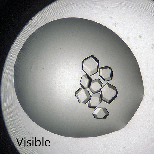
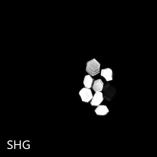
Polarization-resolved second-harmonic generation microscopy as a method to visualize protein-crystal domains
DeWalt EL, Begue VJ, Ronau JA, Sullivan SZ, Das C, Simpson GJ. Polarization-resolved second-harmonic generation microscopy as a method to visualize protein-crystal domains. Acta Crystallographica Section D: Biological Crystallography. 2013;69(Pt 1):74-81. doi:10.1107/S0907444912042503.
Modeling the SHG activities of diverse protein crystals
Haupert LM, DeWalt EL, Simpson GJ. Modeling the SHG activities of diverse protein crystals. Acta Crystallographica Section D: Biological Crystallography. 2012;68(Pt 11):1513-1521. doi:10.1107/S0907444912037638.
Femtosecond nanocrystallography using X-Ray Lasers for membrane protein structure determination
Fromme P, Spence JC. Femtosecond nanocrystallography using X-Ray Lasers for membrane protein structure determination. Current opinion in structural biology. 2011;21(4):509-516. doi:10.1016/j.sbi.2011.06.001.
Selective imaging of active pharmaceutical ingredients in powdered blends with common excipients utilizing two-photon excited ultraviolet-fluorescence and ultraviolet-second order nonlinear optical imaging of chiral crystals
Toth SJ, Madden JT, Taylor LS, Marsac P, Simpson GJ. Selective Imaging of APIs in Powdered Blends with Common Excipients Utilizing TPE-UVF and UV-SONICC. Analytical chemistry. 2012;84(14):5869-5875. doi:10.1021/ac300917t.
2011
Screening of Protein Crystallization Trials by Second Order Nonlinear Optical Imaging of Chiral Crystals (SONICC)
Haupert L, Simpson G. Screening of Protein Crystallization Trials by Second Order Nonlinear Optical Imaging of Chiral Crystals (SONICC). Methods (San Diego, Calif). 2011;55(4):379-386. doi:10.1016/j.ymeth.2011.11.003.
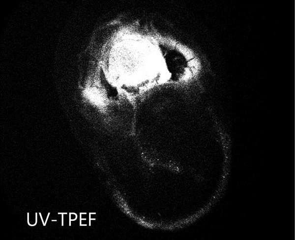
Two-photon excited UV fluorescence for protein crystal detection
Madden JT, DeWalt EL, Simpson GJ. Two-photon excited UV fluorescence for protein crystal detection. Acta Crystallographica Section D: Biological Crystallography. 2011;67(Pt 10):839-846. doi:10.1107/S0907444911028253.
Lipidic cubic phase technologies for membrane protein structural studies
Vadim Cherezov, Lipidic cubic phase technologies for membrane protein structural studies, In Current Opinion in Structural Biology, Volume 21, Issue 4, 2011, Pages 559-566, ISSN 0959-440X, https://doi.org/10.1016/j.sbi.2011.06.007.
Detection of Membrane Protein Two-Dimensional Crystals in Living Cells
Gualtieri EJ, Guo F, Kissick DJ, et al. Detection of Membrane Protein Two-Dimensional Crystals in Living Cells. Biophysical Journal. 2011;100(1):207-214. doi:10.1016/j.bpj.2010.10.051.
2010
Nonlinear optical imaging of integral membrane protein crystals in lipidic mesophases.
Kissick DJ, Gualtieri EJ, Simpson GJ, Cherezov V. Nonlinear Optical Imaging of Integral Membrane Protein Crystals in Lipidic Mesophases. Analytical chemistry. 2010;82(2):491-497. doi:10.1021/ac902139w.
2008
Selective Detection of Protein Crystals by Second Harmonic Microscopy
J. Am. Chem. Soc., 2008, 130 (43), pp 14076–14077
DOI: 10.1021/ja805983b